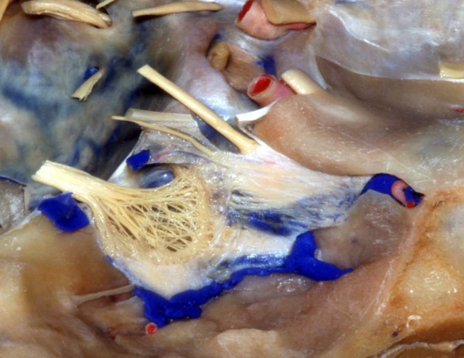
The internal carotid artery and the abducens nerve pass through the cavernous sinus. On its lateral wall from above downwards lie the oculomotor, trochlear and ophthalmic nerves (Fig. 7.60). The maxillary division of the trigeminal goes through the lower part of the lateral wall or just outside the sinus.
What is the structure of the cavernous sinus?
Mar 20, 2022 · The cavernous sinus contains the internal carotid artery and several cranial nerves. Abducens nerve (CN VI) traverses the sinus lateral to the internal carotid artery. The remainder of the cranial nerves pass through the lateral wall of the carotid sinus, and from superior to inferior they are: Oculomotor nerve (CN III) Trochlear nerve (CN IV)
What cranial nerves travel through the cavernous sinus?
Jul 26, 2021 · Structure and Function. The cavernous sinus works as a conduit. Cranial nerves leaving the brainstem travel through the cavernous sinus before entering the orbit to innervate extraocular and intrinsic eye muscles. Also, different venous tributaries drain …
Where does the cavernous sinus receive venous drainage?
Oct 18, 2018 · Travels through cavernous sinus: Travels through lateral wall of cavernous sinus: Abducens nerve (CN VI) Carotid plexus (post-ganglionic sympathetic nerve fibres) Internal carotid artery (cavernous portion) Oculomotor nerve (CN III) Trochlear nerve (CN IV) Ophthalmic (V1) and maxillary (V2) branches of the trigeminal nerve
Where does CN III enter the cavernous sinus?
In addition, the ophthalmic and dorsal meningeal arteries arose from the carotid artery within the cavernous sinus in 8% and 6%, respectively. The three main branches of the meningohypopyseal trunk were the tentorial artery, present in 100%, the dorsal meningeal (90%), and the inferior hypophyseal (80%).
What nerves go through cavernous sinus?
The nerves of the cavernous sinus are the oculomotor nerve (CN III), trochlear nerve (CN IV), ophthalmic nerve (V1), maxillary nerve (V2), abducens nerve (CN VI), and the sympathetic plexus around the internal carotid artery.
Which vessel runs through the cavernous sinus?
internal carotid arteryThe internal carotid artery with its surrounding sympathetic plexus passes through the cavernous sinus. The third, fourth, and sixth cranial nerves are attached to the lateral wall of the sinus. The ophthalmic and maxillary divisions of the fifth cranial nerve are embedded in the wall, as depicted in the image below.
What does not pass through the cavernous sinus?
Trigeminal nerve Prior to entering the cavernous sinus, the proximal portion of the nerve lies in Meckel's cave, where it forms the trigeminal ganglion. After leaving the cave, the mandibular division (CN V3) courses inferiorly to pass through foramen ovale (without entering the cavernous sinus).
What are the contents of the cavernous sinus?
olfactory nerve (CN I)optic nerve (CN II) optic chiasm. ... oculomotor nerve (CN III)trochlear nerve (CN IV)trigeminal nerve (CN V) (mnemonic) trigeminal ganglion.abducens nerve (CN VI)facial nerve (CN VII) (segments mnemonic | branches mnemonic) geniculate ganglion. ... vestibulocochlear nerve (CN VIII)More items...•Jul 31, 2019
Where does the cavernous sinus sit?
The cavernous sinus is located on either side of the pituitary fossa and body of the sphenoid bone between the endosteal and meningeal layers of the dura. It spans from the apex of the orbit to the apex of the petrous temporal bone.Dec 8, 2021
How do facial veins communicate with cavernous sinuses?
The ophthalmic veins drain into the anterior part of the sinus. Emissary veins passing through the foramina in the middle cranial fossa connect the cavernous sinus to the pterygoid plexus of veins and to the facial veins. The superficial middle cerebral vein drains into the cavernous sinus from above.
Where is the petrosal sinus?
temporal boneThe superior petrosal sinus is a small, narrow dural venous sinus found within the anterolateral margin of the tentorium cerebelli. It spans from the cavernous to the transverse sinus by coursing through a shallow groove on the superior border of the petrous part of the temporal bone.Jul 28, 2020
Is the cavernous sinus filled with blood?
It can be life-threatening. The cavernous sinuses are hollow spaces located under the brain, behind each eye socket. A major blood vessel called the jugular vein carries blood through the cavernous sinuses away from the brain.
Where are cavernous sinuses located?
The cavernous sinuses are located within the middle cranial fossa, on either side of the sella turcica of the sphenoid bone (which contains the pituitary gland). They are enclosed by the endosteal and meningeal layers of the dura mater.
Which veins drain into the cavernous sinus?
Each cavernous sinus receives venous drainage from: Ophthalmic veins (superior and inferior) – these enter the cavernous sinus via the superior orbital fissure. Central vein of the retina – drains into the superior ophthalmic vein, or directly into the cavernous sinus.
Overview
The cavernous sinus constitutes a component of dural venous sinus within the brain and consist of multiple neurovasculatures. It is located bilaterally to the sella turcica, extending to the petrous part of the temporal bone in the posterior region, and the superior orbital fissure in the anterior part. It is approximately 2 cm long and 1 cm wide.
Structure
The cavernous sinus acts as a conduit. Before entering the orbit to innervate the eye muscles (both extraocular and intrinsic muscles) the cranial nerves travel through the cavernous sinus once they have left the brainstem. Additionally, drainage of several venous tributaries takes place into the cavernous sinus.
Function
The cavernous sinus is a venous sinus that receives blood from veins including the superior and inferior ophthalmic veins and superficial cortical veins. Typically, it works as a conduit where two important channels drain. This includes the superior and inferior petrosal sinuses draining into the IJV through the sigmoid sinus.
Neurovascular Supply
The bifurcation of the common carotid artery occurs in the cervical region giving rise to the internal and external carotid arteries. Superiorly, the internal carotid artery travels and via the carotid canal enters the skull.
Clinical Relevance and Associated Diseases
Cavernous sinus syndrome is a life-threatening emergency with a wide spectrum of clinical symptoms depending on the target structure that is affected. Any severe lesion that damages the cavernous sinus entirely will result in complete ophthalmoplegia as a result of injury to cranial nerves III, IV and VI.
Which nerve passes through the superior orbital fissure?
On exiting the cavernous sinus, the trochlear nerve passes through the superior orbital fissure into the orbit. The abducens nerve ascends from the lower pons en closed in the dura of the clivus, and passes beneath the petroclinoid (Gruber) ligament within Dorello canal.
Where is the abducens nerve located?
Piercing the dura of the cavernous sinus about 2 cm below the posterior clinoid, the abducens nerve lies freely within the sinus closely adherent to the lateral side of the intracavern ous carotid, and travels through the superior orbital fissure, beneath the oculomotor and trochlear nerves, into the orbit.
What are the two types of sinuses?
1) Superior Petrosal Sinus. 2) Inferior Petrosal Sinus. 3) Foramen Ovale, and other skull base foramina to the pterygoid venous plexus. 4) Contralateral Cavernous sinus thru intercavernous channels. 5) Clival (basilar) venous plexus down to foramen magnum region, and from there into jugular veins or marginal sinus.
What is the basal vein?
The basal vein, in its full expression, is an unbroken conduit between the cavernous sinus and the vein of Galen (see Deep Venous System page for dedicated info). Below is an example of a fully contiguous basal vein (purple), with its deep sylvian component (pink) draining into the cavernous sinus (white) near the confluence of the superficial sylvian vein (blue). The main outflow of the cavernous sinus is the inferior petrosal sinus (black).
Can veins be connected?
yes, all veins are connected. And no, you cant see all the veins that well by injecting arteries. Try injecting veins to see veins. The scalp also drains into the cavernous sinus — we just dont have too many scalp infections yielding cavernous thrombophlebitis to have this fact taught in med school. Here is an example of a scalp AVM draining via both superior ophthalmic veins into the cavernous sinus — case courtesy Eytan Raz MD PhD. The catheter is in the superficial temporal vein. No labels.
Do sylvian veins drain into the cavernous sinus?
The debate as to whether the sylvian veins drain directly into the cavernous sinus or by way of the “sphenoparietal sinus” or Brechet is, in my opinion, won by those who believe in direct venous drainage. A sinus does appear to exist along the sphenoid ridge, and is developmentally related to the middle meningeal veins which are called “anterior parietal”, for good embyologic reasons though they in fact are in the frontal bone. That sinus is likely not connected to the Sylvian veins. In many instances, also, several superficial sylvian veins seem to course in parallel to join the cavernous sinus individually. Either these represent multiple sphenoparietal sinuses, or perhaps all but one of them are true veins, or perhaps all are in fact veins and there is no sphenoparietal sinus related to these. The latter seems to be the preferred option. In the following example, three separate superficial Sylvian veins (purple) run adjacent to the sphenoid ridge to join a common channel (white) which may be a common vein or perhaps a short sphenoparietal sinus.
Is the cavernous sinus a metaphysical entity?
Cavernous Sinus. The cavernous sinus is, at least from the angiographic perspective, a metaphysical entity. It is a collection of extradural venous compartments, often functionally separate, which altogether constitute the venous space we have come to regard as a distinct anatomical structure. It is critical for the neurointerventionalist ...
What is the cavernous sinus?
The cavernous sinus within the human head is one of the dural venous sinuses creating a cavity called the lateral sellar compartment bordered by the temporal bone of the skull and the sphenoid bone, lateral to the sella turcica .
Where is the cavernous sinus located?
Cavernous sinus (center, labeled "SIN. CAVERN.") The sinuses at the base of the skull. Cavernous sinus labeled in red. The cavernous sinus within the human head is one of the dural venous sinuses creating a cavity called the lateral sellar compartment bordered by the temporal bone of the skull and the sphenoid bone, lateral to the sella turcica .
Where is the pituitary gland located?
The pituitary gland lies between the two paired cavernous sinuses. An abnormally growing pituitary adenoma, sitting on the bony sella turcica, will expand in the direction of least resistance and eventually invade the cavernous sinus.
What is the cause of cavernous sinus syndrome?
Cavernous sinus syndrome may result from mass effect of these tumors and cause ophthalmoplegia (from compression of the oculomotor nerve, trochlear nerve, and abducens nerve), ophthalmic sensory loss (from compression of the ophthalmic nerve), and maxillary sensory loss (from compression of the maxillary nerve).
What happens if the internal carotid artery ruptures?
Lesions affecting the cavernous sinus may affect isolated nerves or all the nerves traversing through it.
Overview
Structure
- The cavernous sinus acts as a conduit. Before entering the orbit to innervate the eye muscles (both extraocular and intrinsic muscles) the cranial nerves travel through the cavernous sinus once they have left the brainstem. Additionally, drainage of several venous tributaries takes place into the cavernous sinus. Venous blood from the central retin...
Function
- The cavernous sinus is a venous sinus that receives blood from veins including the superior and inferior ophthalmic veinsand superficial cortical veins. Typically, it works as a conduit where two important channels drain. This includes the superior and inferior petrosal sinuses draining into the IJV through the sigmoid sinus. In addition, drainage with emissary vein…
Neurovascular Supply
- The bifurcation of the common carotid artery occurs in the cervical region giving rise to the internal and external carotid arteries. Superiorly, the internal carotid artery travels and via the carotid canal enters the skull. Following this, the internal carotid artery enters the carotid canal and makes a 90-degree turn, travelling in the horizontal direction in the petrous part of the temp…
Clinical Relevance and Associated Diseases
- Cavernous Sinus Syndrome
Cavernous sinus syndrome is a life-threatening emergency with a wide spectrum of clinical symptoms depending on the target structure that is affected. Any severe lesion that damages the cavernous sinus entirely will result in complete ophthalmoplegia as a result of injury to cranial n… - Cavernous Sinus Infections
Blood from the facial vein may also drain to the cavernous sinus in certain cases promoting a pathway for infection. This may cause the infection to spread from the face to the cavernous sinus, and from there to the brain. The content shared in the Health Literacy Hub website is provi…
Popular Posts:
- 1. which institute is best for medical transcription course in bangalore
- 2. what is financial management course
- 3. what is the best lsat prep course to take
- 4. how to write a college course curriculum
- 5. how long is billing and coding course
- 6. how long is medical assistant course
- 7. what is pmp training course
- 8. what is a course management system
- 9. what is business management course
- 10. how long is hvac course