For the most part, the spinal nerves exit the vertebral canal through the intervertebral foramen below their corresponding vertebra. Therefore, there are 12 pairs of thoracic spinal nerves, 5 pairs of lumbar spinal nerves, 5 pairs of sacral spinal nerves, and a coccygeal nerve.
What are spinal nerves?
The spinal nerves are relatively large nerves that are formed by the merging of a sensory nerve root and a motor nerve root. These nerve roots emerge directly from the spinal cord—sensory nerve roots from the back of the spinal cord and the motor nerve roots from the front of the spinal cord.
How do nerves work in the cervical spine?
These complex networks of nerves enable the brain to receive sensory inputs from the skin and to send motor controls for muscle movements. In the cervical spine, there are eight pairs of spinal nerves labeled C1 to C8, which innervate the neck, shoulder, arm, hand, and more. Video: Cervical Nerve Anatomy
How are spinal nerves named according to where they exit?
Again, they are named according to where they each exit in the spine (see figure below). Each spinal nerve is attached to the spinal cord by two roots: a dorsal (or posterior) root which relays sensory information and a ventral (or anterior) root which relays motor information.
Where do the spinal nerves receive sensory messages?
The spinal nerves receive sensory messages from tiny nerves located in areas such as the skin, internal organs, and bones. The spinal nerves send sensory messages to the sensory roots, then to sensory fibers in the posterior (back or dorsal) part of the spinal cord.
Where do the spinal nerves come from?
These nerve roots emerge directly from the spinal cord—sensory nerve roots from the back of the spinal cord and the motor nerve roots from the front of the spinal cord. As they join, they form the spinal nerves on the sides of the spinal cord.
What are the nerves that connect the spinal cord to the body?
The spinal nerves are peripheral nerves that transmit messages between the spinal cord and the rest of the body, including muscles, skin, and internal organs. Each spinal nerve is dedicated to certain regions of the body.
What causes a pinched nerve in the spine?
A pinched nerve occurs when there is pressure or compression of a spinal nerve , and it is the most common spinal nerve disorder.
How to diagnose spinal nerve problems?
The first is a physical examination, which can identify impairment corresponding to a dermatome and/or myotome. Reflexes also correspond to spinal nerves, and they are usually diminished in these situations as well, further helping to identify which nerves are involved.
What are the major nerves in the body?
Spinal nerves are the major nerves of the body. A total of 31 pairs of spinal nerves control motor, sensory, and other functions. These nerves are located at the cervical, thoracic, lumbar, sacral, and coccygeal levels.
Why is the spinal nerve function impaired?
In these instances, the spinal nerve function is impaired because the nerve fibers in the nearby sections of the spine cease to send or receive messages to and from the spinal nerves. Treatment of spine disease depends on the cause.
Which spinal nerves are involved in the plexus?
There are five main plexi formed by the spinal nerves: Cervical Plexus: Composed of the merging of spinal nerves C1 through 5, these divide into smaller nerves that carry sensory messages and provide motor control to the muscles of the neck and shoulders.
What is the function of the spinal nerve?
Therefore, once the two roots come together to form the spinal nerve, the nerve carries a combination of both sensory and motor information (i.e.
Where do spinal nerves exit?
they are named in accordance with the level of the spine they exit from. E.g. the C2 nerve exits between the C1-2 vertebrae, the L4 nerve exits between the L4-5 vertebrae.
What is the difference between cervical plexus and brachial plexus?
Cervical Plexus – the cervical plexus represents the continuation of the upper cervical spinal nerves that gives innervation (i.e. supplies nerve function) to the neck and shoulders. Brachial Plexus – the brachial plexus represents the continuation of the lower cervical spinal nerves that gives innervation to the upper back, shoulder, arms, ...
How many nerves are in the peripheral nervous system?
The Peripheral Nervous System (PNS) consists of 12 cranial nerves, and 31 pairs of spinal nerves. The PNS acts as the system of electrical wires that allows for communication between the CNS and the body’s muscles and sensory receptors. They also control the automatic functions of the bowel, bladder, respiratory (breathing), and heart function.
How many categories of spinal nerves are there?
The spinal nerves are divided into four main categories of spinal nerves based on the location from which they branch
Which nerves give innervation to the lower extremities?
Lumbar Plexus – the lumbar plexus represents the continuation of lumbar spinal nerves that give innervation to the lower extremities. Sacral Plexus – the sacral plexus gives innervation to the back of the thigh, leg, bottom of the foot, as well as the pelvis.
Where do the lumbar nerves come from?
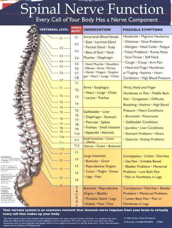
5 lumbar (L1-L5) nerves emerge from the lumbar spine (lower back) 5 sacral (S1-S5) nerves emerge from the sacrum (the triangular bone at the base of the spine) 1 coccygeal nerve emerges from the coccyx (the tailbone) Below is a chart that outlines the main functions of each of the spine nerve roots: Spinal Nerve Root.
Where do spinal nerves come from?
Spinal nerves emerge from the spinal column through an opening between adjacent vertebrae (known as intervertebral foramen). This is the case for all of the spinal nerves except the first pair, which emerge between the occipital bone and the uppermost vertebrae.
Which nerves are distributed evenly along the spinal cord and the spine?
Spinal nerves are relatively large nerves which are distributed evenly along the spinal cord and the spine. The spine is a column of vertebrae bones which protects the spinal cord.
How many spinal nerves are there?
These nerves are an integral part of the PNS in that they control motor, sensory, and autonomic functions between the spinal cord and the body. There are 31 pairs of spinal nerves, located at the cervical, thoracic, lumbar, sacral, and coccygeal levels:
What is the name of the condition that attacks the myelin sheath of the spinal cord?
Guillan Barre Syndrome (GBS) – this is a condition which attacks the myelin sheath (protective insulating layer) of the neurons. As this is a demyelinating condition, it can weaken the spinal nerves over time and cause weakness and tingling sensations all over the body. Eventually, it may even impair the muscles which control breathing.
How many nerve rootlets are there in the spinal cord?
Each nerve root comprises of approximately 8 nerve rootlets and as they join together, they form the spinal nerves which project off the spinal cord.
What causes pain and inflammation in the spine?
Spinal infections – types of spinal infections can include disc infections and spinal bone infections and typically cause inflammation and pain which may travel into other parts of the body. A spinal infection may begin near spinal nerve roots and thus will take effect on the spinal nerves which branch from it.
What is the condition where the spinal cord is compressed?
Compressive neuropathy – this condition can occur when the spinal nerves are compressed. This can happen when the nerves that exit the spinal cord become trapped or swollen and it can be extremely painful.
What is the spinal nerve?
They are the conduits in the relay of information that occurs between the brain and peripheral tissues. Sensory information relays from the peripheral tissues into the brain via the dorsal root. Motor information relays from the brain to the muscles in the peripheral tissues via the ventral root.
How do spinal nerves connect to the spine?
The spinal nerves connect to the spine via dorsal and ventral roots. The dorsal root contains sensory input and the ventral root contains motor output. They then combine and further bifurcate into dorsal and ventral rami that include both sensory and motor information before projecting towards the peripheral tissues they innervate.
How many thoracic nerves are there?
There are 12 total thoracic spinal nerve pairs termed T1-T12. They innervate the chest and the torso, in addition to providing aid with functioning of organs in the chest and torso. The intercostal nerves emerge from T1-T11 whereas the subcostal nerve emerges from T12. The posterior division of thoracic nerves provide motor innervation to rhomboid and trapezius muscles in the back.
What is the name of the group of nerve bundles that innervate the same region?
Nerve plexuses are groups of nerve bundles that innervate the same bodily region. Unlike spinal nerves, nerve plexuses are formed with combination of spinal nerves from different section of the spine. Thus, they are classified by their function and named after the body region they innervate. The exception to this is the Auerbach plexus, which innervates the GI tract and is named after the person who discovered it. The most common plexuses are known as:
How many cervical spinal nerves are there?
There are 8 total cervical spinal nerves termed C1-C8. They innervate the head and neck and the arms. They provide motor innervation to the three muscles in the neck called sternohyoid, sternothyroid, and omohyoid muscles. Some of the cervical nerves have specific names as described below:
What is the name of the nerve plexus?
A nerve plexus is a network of nerves that innervate the same region of the body. They are typically named after the regions of the body they innervate also, such as the cervical, brachial, lumbar, sacral, celiac and coccygeal plexus. Auerbach's plexus is located in the GI tract. Unlike spinal nerve divisions, nerve plexuses are not limited to branches from same sections of the spinal cord; instead, nerve plexuses are often combinations of spinal nerves from different parts of the spine. For example, the brachial plexus is formed by C5-T1 spinal nerves. Thus, nerve plexuses are classified based on their function. The most common nerve plexuses are:
How many nerves are there in the spine?
From the space between each vertebrae, a pair of nerves projects outwards on both sides of the spine. These 31 pairs of nerves are collectively defined as the spinal nerves. They connect to the spine via a dorsal (back) root and a ventral (front) root which then combines before again bifurcating into dorsal and ventral rami that project to the tissues they innervate. The dorsal root contains sensory or afferent input from the tissues to the brain, whereas the ventral root carries motor or efferent output the tissue from the brain.
How many nerves are in the spinal cord?
In the thoracic region of the spinal cord, we have 12 pairs of nerves that attach, one on either side, of course. And then both in the lumbar region And in the sacral region of the cord we have five nerves that attach, five lumbar and five sacral.
How many spinal nerves are there?
So there are 31 pairs of spinal nerves. eight cervical, 12 thoracic, five lumbar, five sacral, and one coccygeal. Now, that's one way of characterizing the long axis of the spinal cord. Another way is to note that there are, two regions where the diameter of the spinal cord is enlarged.
What is the space between the vertebrae called?
From the lumbosacral enlargement out through the canals between the vertebrae. space called the anterior vertebral foramina, that allow for the passage of the spinal nerve routes.
How many pairs of spinal nerves are in the cervical region?
By the attachment of 31 pairs of spinal nerves. So, in the cervical region of the spinal cord, we have eight pairs of nerves that attach.
What is medical neuroscience?
Medical Neuroscience explores the functional organization and neurophysiology of the human central nervous system, while providing a neurobiological framework for understanding human behavior. In this course, you will discover the organization of the neural systems in the brain and spinal cord that mediate sensation, motivate bodily action, ...
Where does the spinal nerve travel?
The spinal nerve travels a short distance inside the intervertebral foramen, after which it branches off into several nerves that innervate different parts of the body. Doctors may sometimes refer to the part of the spinal nerve exiting the intervertebral foramen as the nerve root or use the terms nerve root and spinal nerve interchangeably.
What are the two nerves that branch off from the right and left sides of the spinal cord?
Lumbar Spinal Nerves. Two spinal nerves branch off from the right and left sides of the spinal cord or the cauda equina at each spinal segment. These spinal nerves are formed by 2 types of fibers—sensory fibers that send messages to the brain (feeling pain when the leg is hurt) and motor fibers that receive messages from the brain ...
How many pairs of lumbar nerves innervate the lower limbs?
The 5 pairs of lumbar spinal nerves innervate the lower limbs. While innervation can vary among individuals, some common patterns include 2:
How many pairs of lumbar spinal nerves are there?
There are 5 pairs of lumbar spinal nerves that progressively increase in size from L1 to L5. These nerves exit the intervertebral foramina below the corresponding vertebra. For example, the L4 nerve exits beneath the L4 vertebra through the L4-L5 foramen. These nerves course down from the lower back and merge with other nerves to form ...
Why do lumbar nerves get compressed?
1 This anatomy, in addition to lower back disorders, such as disc herniation or degeneration may cause the nerve to get compressed, resulting in leg pain and weakness.
What nerves are involved in the back of the leg?
The L4 and L5 nerves (along with other sacral nerves) contribute to the formation of the large sciatic nerve that runs down from the rear pelvis into the back of the leg and terminates in the foot. Symptoms and signs arising from these nerves, typically referred to as sciatica, can cause a sharp, burning pain radiating down the leg with associated weakness and numbness.
What nerve controls the hip, knee, foot, and toe?
The L5 spinal nerve controls hip, knee, foot, and toe movements. Read more about Spinal Cord and Spinal Nerve Roots. advertisement.
Which nerves branch off from the spinal cord?
Cervical Spinal Nerves. Spinal nerves branch off from the spinal cord to innervate the rest of the body. These complex networks of nerves enable the brain to receive sensory inputs from the skin and to send motor controls for muscle movements. In the cervical spine, there are eight pairs of spinal nerves labeled C1 to C8, which innervate the neck, ...
How many pairs of nerves are there in the cervical spine?
In the cervical spine, there are eight pairs of spinal nerves labeled C1 to C8, which innervate the neck, shoulder, arm, hand, and more.
What happens if a cervical nerve is compressed?
If a cervical nerve becomes irritated or compressed, it may cause pain and/or dysfunction that correlates to its dermatome for sensations and/or myotome for motor control. See Spinal Cord Compression and Dysfunction from Cervical Stenosis.
What is the name of the nerve that connects the ventral and dorsal roots?
When the ventral and dorsal roots merge, the combined nerve is called the spinal nerve. From there, the spinal nerve branches into a network ...
What is the function of the cervical nerve?
Cervical spinal nerves, also called cervical nerves, provide functional control and sensation to different parts of the body based on the spinal level where they branch out from the spinal cord. While innervation can vary from person to person, some common patterns include:
Which nerve root innervates the specific region of skin that it covers?
Dorsal root (located in back) that carries sensory signals back to the brain from that nerve root’s dermatome, which innervates the specific region of skin that it covers. Watch Cervical Spinal Cord Anatomy Animation. advertisement. The ventral root and dorsal root branch off separately from the spinal cord then merge together in ...
Which cervical nerves control the neck?
C1, C2, and C3 (the first three cervical nerves) help control the head and neck, including movements forward, backward, and to the sides. 1 The C2 dermatome handles sensation for the upper part of the head, and the C3 dermatome covers the side of the face and back of the head. 2 (C1 does not have a dermatome.)
Anatomy

The spine is made up of vertebrae (back bones) that protect and surround the spinal cord, which is a column of nerve tissue. Spinal nerves branch out from the spinal cord. These are peripheral nerves, or those that run through other parts of the body and transmit message to and from the brain/spinal cord. These nerves …
Function
- The spinal nerves have small sensory and motor branches. Each of the spinal nerves carries out functions that correspond to a certain region of the body. These are muscle movement, sensation, and autonomic functions(involuntary functions).
Associated Conditions
- Spinal nerves can be affected by a number of conditions. These situations can cause pain, sensory changes, and/or weakness. The diagnosis of a spinal nerve problem involves several steps. The first is a physical examination, which can identify impairment corresponding to a dermatome and/or myotome. Reflexes also correspond to spinal nerves, and they are usually di…
Rehabilitation
- Most of the time, spinal nerve impairment is treatable. Mild inflammation can usually be managed with anti-inflammatory medication, and pain can usually be lessened with over-the-counter pain relievers. Physical therapy and exercises can help alleviate pressure and improve posture and muscle tone, reducing pain. However, pain can be persistent and severe, requiring more aggress…
Summary
- There are 31 pairs of spinal nerves that branch out from the spinal cord. Each carries out functions that correspond to a certain region of the body, Many spine-related diseases, viral infections, and traumatic injuries can affect spinal nerves and lead to pain, weakness, and/or loss of sensation. Treatments for spinal nerve impairment depend on the cause, but a full or partial r…
Popular Posts:
- 1. which institute is best for medical transcription course in bangalore
- 2. what is financial management course
- 3. what is the best lsat prep course to take
- 4. how to write a college course curriculum
- 5. how long is billing and coding course
- 6. how long is medical assistant course
- 7. what is pmp training course
- 8. what is a course management system
- 9. what is business management course
- 10. how long is hvac course