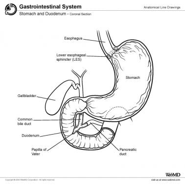
The pancreas, liver and gallbladder all deliver their digestive secretions into the duodenum through an orifice known as the ampulla of Vater, which is located roughly in the middle of the duodenum on the left side.
Full Answer
What blood vessels supply blood to the duodenum?
Blood Supply. The supply of blood to the duodenum is carried by the anterior and posterior superior pancreaticoduodenal arteries (branches of the gastroduodenal artery) and the inferior pancreaticoduodenal artery (branch of the superior mesenteric artery) which form an arterial arcade.
What is the lining of the duodenum composed of?
The lining of the duodenum is composed of four layers—each with its own specialized function. The duodenum measures approximately 20 to 25 centimeters (approximately 8 to 10 inches) in length (compared with the jejunum, which is approximately 2.5 meters, or 8 feet, long). 1
What is the venous drainage of the duodenum?
The correspondent veins are responsible for the venous drainage. The sympathetic innervation is carried by nerves of the coeliac plexus, the parasympathetic innervation by the vagus nerve (cranial nerve X). For more details about the duodenum, take a look at the following resources:
How is the duodenum connected to the liver?
The first segment of the duodenum —the superior part of the duodenum (called the duodenal bulb) is connected to the liver via the hepatoduodenal ligament. This connection allows for transportation of nutrients from the small intestine to the liver; it also allows the duodenum to receive bile from the liver.
What blood vessels supply the duodenum?
The proximal segment of the duodenum is supplied by the gastroduodenal artery and its branches which include the superior pancreaticoduodenal artery. The distal segment of the duodenum is supplied by the superior mesenteric artery and the inferior pancreaticoduodenal artery.
What artery runs behind the duodenum?
Gastroduodenal ArteryThe gastroduodenal artery arising from the common hepatic artery, a branch of the celiac trunk, becomes the superior pancreaticoduodenal artery as it passes behind the first portion of the duodenum.
What artery passes posterior to the duodenum?
The gastroduodenal artery arises from the common hepatic artery posterosuperior to the upper border of the superior part of the duodenum. It courses inferiorly behind the duodenum and terminates at its lower border, where it divides into its terminal branches.
Which vessels enter the duodenum at the duodenal papilla?
The pancreatic duct and common bile duct enter the descending duodenum through the major duodenal papilla (ampulla of Vater).
What artery supplies the stomach and duodenum?
The celiac artery gives rise to three major branches, including the left gastric, splenic, and common hepatic arteries. Collectively, these major branches of the celiac artery supply the stomach, spleen, liver, gallbladder, abdominal esophagus, pancreas, and duodenum.
Where is the SMA artery?
In human anatomy, the superior mesenteric artery (SMA) arises from the anterior surface of the abdominal aorta, just inferior to the origin of the celiac trunk, and supplies blood to the intestine from the lower part of the duodenum through two-thirds of the transverse colon, as well as the pancreas.
Where does celiac artery come from?
The celiac trunk, also known as the celiac artery, is a short vessel that arises from the aorta and passes below the median arcuate ligament, just as the aorta enters the abdomen at the level of the T12 vertebra. The celiac trunk measures about 1.5cm to 2cm in length.
Where does the Supraduodenal artery come from?
The supraduodenal artery (SDA) is a branch of the gastroduodenal artery (GDA). It arises soon after the origin of the GDA posterior to the first part of the duodenum and supplies the anterosuperior part of the first and second parts of the duodenum, contributing to the rich arterial anastomotic supply of the duodenum.
What are mesenteric vessels?
The mesenteric arteries take blood from the aorta and distribute it to a large portion of the gastrointestinal tract.
Where is ligament of Treitz located?
Where is the ligament of Treitz? The ligament of Treitz extends from the diaphragm to a part of the small intestine called the duodenojejunal flexure. The duodenojejunal flexure is a sharp angle in the small intestine between the duodenum and the jejunum (two parts of the small intestine).
What does the ligament of Treitz connect?
The suspensory muscle of duodenum is a thin muscle connecting the junction between the duodenum, jejunum, and duodenojejunal flexure to connective tissue surrounding the superior mesenteric artery and coeliac artery. It is also known as the ligament of Treitz.
What is Treitz ligament?
The ligament of Treitz is one of the frequently forgotten structures within the abdomen. It was named after the Austrian physician and anatomist Wenzel Treitz, who in 1853 first described the ligament as a thin, triangular, fibromuscular band extending from the upper surface of the duodenojejunal junction [1].
Which artery supplies blood to the duodenum?
The supply of blood to the duodenum is carried by the anterior and posterior superior pancreaticoduodenal arteries (branches of the gastroduodenal artery) and the inferior pancreaticoduodenal artery (branch of the superior mesenteric artery) which form an arterial arcade.
What is the duodenum?
Histology. Histologically the duodenum is similar to all the other hollow organs of the gastrointestinal tract: mucosa, submucosa and muscular is. The mucosa consists of simple columnar epithelium ( lamina epithelialis ), a connective tissue layer ( lamina propria) and a smooth muscle layer ( lamina muscularis ).
What are the structures that increase the absorption area of the duodenum?
Typical for all sections of the small intestines are microvilli (hairlike structures projecting from the surface), finger-shaped villi and circular folds of the mucosa and submucosa (valves of Kerckring). These structures increase the absorption area of the duodenum up to 1500 times.
What part of the small intestine is responsible for digesting food?
Last reviewed: June 17, 2021. Reading time: 6 minutes. The duodenum is the first of the three parts of the small intestine that receives partially digested food from the stomach and begins with the absorption of nutrients. It is directly attached to the pylorus of the stomach. It has a C-shape, it is closely related to the head ...
What are the characteristics of the duodenum?
A characteristic feature of the duodenum is the Brunner’s glands embedded in the submucosa. These produce – amongst others – mucous secret containing bicarbonate which serves to neutralize the gastric acid. Furthermore crypts of Lieberkuhn lie between the villi. Paneth cells are found in the lumen of these crypts.
Which part of the liver is connected to the liver?
The superior part (first part, D1) lies intraperitoneally and is enlarged proximally ( duodenal bulb ). It is connected to the liver by the hepatoduodenal ligament. The superior part ends at the superior duodenal flexure and becomes the descending part.
Which organ is easy to locate during a dissection?
The duodenum follows a C-shaped trajectory around the head of the pancreas.
What is the second part of the duodenum?
Descending (second) part. It extends from superior duodenal flexure (L1) to inferior duodenal flexure (L3). The main features of the second part of the duodenum are; Its upper half develops from the foregut and the lower half from the midgut. It lies behind the transverse mesocolon.
Which layer of the duodenum contains connective tissue cells?
Serosa (visceral peritoneum): It contains connective tissue cells, blood vessels, and adipose cells. The outermost layer of the first part of the duodenum.
What are the relations of the ascending (fourth) part of the duodenum?
The relations of ascending (fourth) part of the duodenum is: Anteriorly: Transverse colon and mesocolon. Posteriorly: Left psoas major muscle, left sympathetic chain, left gonadal vessels, and inferior mesenteric vein (IVC). Superiorly: Body of the pancreas. On to the left: Left side kidney and left ureter.
What is the shortest part of the small intestine?
The duodenum is the shortest, broadest (widest), and most fixed part of the small intestine. It extend from the pylorus to duodenojejunal flexure. The length of the duodenum is about 10 inches (25 cm) long.
Where does the pylorus of the stomach start?
It starts at the pylorus of the stomach which lies on the transpyloric plane around 2.5 cm to the right of the median plane & ends at the duodenojejunal junction which lies around 2.5 cm to the left of the median plane and a little beneath the transpyloric plane.
Which muscle is attached to the right crus of the diaphragm?
Suspensory muscle of the duodenum (LIGAMENT OF TREITZ) It is a fibromuscular band, which suspends the duodenojejunal flexure from the right crus of the diaphragm. Its upper end is attached to the right crus of the diaphragm and the lower end attached to the posterior surface of the duodenojejunal flexure.
What is the importance of the duodenum?
The main significance of the duodenum is the digestion of the food. It is C- shaped structure, near the head of the pancreas in the human. It located in the abdominal cavity above the level of the umbilicus; opposite the first (L1), second (L2), and third (L3) lumbar vertebrae.
What is the duodenum?
Located inferior to the stomach, the duodenum is a 10-12 inch (25-30 cm) long C-shaped, hollow tube. The duodenum is a part of the gastrointestinal (GI) tract, attached to the pyloric sphincter of the stomach on its superior end and to the jejunum of the small intestine on its inferior end. Continue Scrolling To Read More Below...
Which layer of the duodenum contains smooth muscle tissue?
Many blood vessels and nerves pass through the submucosa, while protein fibers give strength and elasticity to the duodenum. Surrounding the submucosa is the muscularis layer that contains the smooth muscle tissue of the duodenum. Contractions of the muscularis mix chyme and propel it through the duodenum toward the rest of the small intestine.
Which part of the small intestine is responsible for the digestion of food?
Duodenum. The duodenum is the first and shortest segment of the small intestine. It receives partially digested food (known as chyme) from the stomach and plays a vital role in the chemical digestion of chyme in preparation for absorption in the small intestine.
Which glands secrete alkaline mucus?
Next, Brunner’s glands in the mucosa of the duodenum secrete alkaline mucus containing a high concentration of bicarbonate ions to neutralize the hydrochloric acid present in the chyme. This alkaline mucus both protects the walls of the duodenum and helps the chyme to reach a pH conducive to chemical digestion in the small intestine.
Which layer of the duodenum is the outermost layer of the intestine?
Lastly, the serosa is the outermost layer of the duodenum that acts as the outer skin of the intestine. Serous membrane made of simple squamous epithelium provides a smooth, slick surface to prevent friction between the duodenum and the surrounding organs. The serosa also secretes serous fluid to further reduce friction and keep ...
Where does peristalsis flow?
Slow waves of smooth muscle contraction known as peristalsis flow down the length of the gastrointestinal tract to push chyme through the duodenum. Each wave begins at the stomach and pushes chyme a short distance toward the jejunum.
What is the function of bile in the liver?
Bile produced in the liver and stored in the gallbladder acts as an emulsifier, breaking lipids into smaller globules to increase their surface area. Pancreatic juice contains many enzymes to break carbohydrates, lipids, proteins and nucleic acids into their monomer subunits.
Which artery supplies the duodenum?
Above the level of opening of bile duct, duodenum is supplied by superior pancreatico-duodenal artery and below by inferior pancreatico-duodenal artery. First part of duodenum receives additional supply from: Right gastric artery. Supraduodenal artery (branch of hepatic artery)
How many parts does the duodenum have?
Duodenum is divided into four parts: Superior (First) part – from pyloric end to superior duodenal flexure (5cm.or 2”). Descending (Second) part – from superior duodenal flexure to inferior duodenal flexure (7.5cm. or 3”) Horizontal (Third) part – from inferior duodenal flexure to the front of aorta (10cm. or 4”) ...
What is the major duodenal papilla?
Major duodenal papilla: is a conical elevation situated 8-10cm from the pyloric end of the stomach. Ampulla of Vater (fromed by union of bile duct and main pancreatic duct) opens on its summit. The opening is guarded by sphincter of Oddi.
What is the horizontal part of the aorta?
Horizontal (Third) part – from inferior duodenal flexure to the front of aorta (10cm. or 4”) Ascending (Fourth) part – from the front of aorta to duodenojejunal flexure (2.5cm or 1”).
What is the condition where the pancreas encircles the duodenum?
Annular pancreas. It is a rare condition in which pancreatic tissue encircles the 2nd part of the duodenum, causing duodenal obstruction It occurs due to abnormal embryological development. It can result from growth of a bifid ventral pancreatic bud around the duodenum.
Which artery is the right gastric artery?
Right gastric artery. Supraduodenal artery (branch of hepatic artery) Retroduodenal artery ( branch of gastroduodenal artery) Right gastroepiploic artery. Duodenum develops from both foregut and midgut, therefore it is supplied by the branches of arteries of foregut (celiac trunk) and midgut (superior mesenteric artery).
What is the shortest part of the small intestine?
Describe the gross features and location of Duodenum. It is the shortest, widest and more or less fixed proximal part of the small intestine. It is 25cm or 10 inches (equivalent to 12 fingers) in length. It extends from the pyloric end of the stomach to duodenal-jejunal flexure.
What is the blood supply of the duodenum?
The blood supply of the C-shaped duodenum is shared with the head of the pancreas. The proximal segment of the duodenum is supplied by the gastroduodenal artery and its branches which include the superior pancreaticoduodenal artery. The distal segment of the duodenum is supplied by the superior mesenteric artery and the inferior pancreaticoduodenal artery. The venous drainage follows the arteries and ultimately drains into the portal system. The duodenum also has lymphatic vessels which drain into the pancreaticoduodenal lymph nodes located along the pancreaticoduodenal vessels and the superior mesenteric lymph nodes.
What is the duodenum?
What is duodenum. The duodenum is the first portion of the small intestine. The duodenum is the initial C-shaped segment of the small intestine and is a continuation of the pylorus (part of stomach) 1). Distally, duodenum is in continuation with the jejunum and ileum, with the proximal segment being the shortest and widest.
What is the mixing pot of the small intestine?
The duodenum is the mixing pot of the small intestine. Duodenum receives chyme from the stomach, which is a mixture of food products and acid, through a controlled valve between the stomach and the duodenum called the pylorus.
What are the 4 segments of the duodenum?
The 4 segments of the duodenum include the following: The duodenal bulb, which connects to the undersurface of the liver via the hepatoduodenal ligament, which contains the portal vein, the hepatic artery, and common bile duct.
Which muscle prevents reflux of duodenal secretions into the bile and pancreatic ducts
This is received into the duodenum by the major and minor papilla in the second part of the duodenum. The duodenal papilla is surrounded by a semicircular fold superiorly and the sphincter of Oddi which is the muscle that prevents reflux of duodenal secretions into the bile and pancreatic ducts.
Where is the duodenojejunal flexure located?
The duodenojejunal flexure is the sudden turn which is usually identified during surgery by the location of the inferior mesenteric vein, which is located to the immediate left. The duodenojejunal flexure is attached to the posterior abdominal wall by the ligament of Treitz. Except for the first segment, the rest of the duodenum is retroperitoneal ...
What nerves travel through the duodenum?
These parasympathetic nerves pass through the celiac plexuses and follow the celiac trunk toward the duodenum. The nerves then synapse in ganglia in the gut plexuses in the duodenum and reach their final targets through short postsynaptic fibers. The sympathetic nerves are branches of the celiac plexus which originate from T5 through T9. These sympathetic nerves pass through the sympathetic chain and travel through the greater splanchnic nerve and synapse in the celiac ganglia. The postsynaptic sympathetic follow the branches of the celiac trunk toward the duodenum.
What is the cross section of the superior duodenum?
A cross section of the superior duodenum, showing the myenteric nerve plexus (*), large clusters of Brunner glands (vertical centre), and a secretory duct (D) extending horizontally from a Brunner gland. The duodenum is often involved in the diseases of its neighbours, in particular the pancreas and the biliary tract.
What is the disease of the duodenum?
Inflammation of the duodenum is known as duodenitis, which has various causes, prominent among them infection by the bacterium Helicobacter pylori.
What causes peptic ulcers in the duodenum?
H. pylori increases the suscept ibility of the duodenal mu cosa to damage from unneutralized digestive acids and is a major cause of peptic ulcers, the most common health problem affecting the duodenum. Other conditions that may be associated with duodenitis include celiac disease, Crohn disease, and Whipple disease.
Where do the duodenal papilla and gallbladder enter?
Ducts from the pancreas and gallbladder enter at the major duodenal papilla (papilla of Vater) in the descending duodenum, bringing bicarbonate to neutralize the acid in the gastric secretions, pancreatic enzymes to further digestion, and bile salts to emulsify fat.
Where does food enter the duodenum?
A liquid mixture of food and gastric secretions enters the superior duodenum from the pylorus of the stomach, triggering the release of pancreas -stimulating hormones (e.g., secretin) from glands (crypts of Lieberkühn) in the duodenal wall.
Why is the duodenum compressed?
The horizontal duodenum, because of its location between the liver, pancreas, and major blood vessels, can become compressed by those structures in people who are severely thin, requiring surgical release to eliminate painful duodenal dilatation, nausea, and vomiting. duodenum. Endoscopic image of the duodenum. Samir.
What are the four segments of the duodenum?
On anatomic and functional grounds, the duodenum can be divided into four segments: the superior (duodenal bulb), descending, horizontal, and ascending duodenum.
Where does the duodenum begin?
The duodenum begins at the duodenal bulb and ends at the ligament of Treitz, where it continues as the jejunum (this is often called the duodenojejunal (DJ) flexure ). It is composed of four distinct parts and is neither wholly peritoneal nor retroperitoneal.
What is the first part of the duodenum?
First part (D1) The first (superior) part of the duodenum begins as a continuation of the duodenal end of the pylorus. It passes laterally to the right, superiorly and posteriorly, for approximately 5 cm, before making a sharp curve inferiorly into the superior duodenal flexure. It is intraperitoneal for the first 2-3 cm only.
What is the first part of the small intestine?
Duodenum. The duodenum (plural: duodena or duodenums) is the first part of the small intestine and is the continuation of the stomach.
Which part of the duodenum is the pancreatic duct?
medially: pancreatic head. The pancreatic duct and common bile duct enter the descending duodenum through the major duodenal papilla ( ampulla of Vater ). This part of the duodenum also contains the minor duodenal papilla, the entrance for the accessory pancreatic duct.
Where is the duodenum located?
Gross anatomy. The duodenum is a 20-30 cm C-shaped hollow viscus predominantly on the right side of the vertebral column. It lies at the level of L1-3 and the convexity of the duodenum (called the duodenal sweep by radiologists) usually encompasses the head of the pancreas. The duodenum begins at the duodenal bulb and ends at the ligament ...
Where is the second part of the duodenum?
Second part (D2) The second (descending) part of the duodenum begins at the superior duodenal flexure. It passes inferiorly to the lower border of vertebral body L3, before making a sharp turn medially into the inferior duodenal flexure, the end of the descending part. Relations 3: anteriorly: transverse mesocolon.
Where does the fourth part of the pancreas curve?
Then, it curves anteriorly and terminates at the duodenojejunal flexure where it joins the jejunum.
Popular Posts:
- 1. which institute is best for medical transcription course in bangalore
- 2. what is financial management course
- 3. what is the best lsat prep course to take
- 4. how to write a college course curriculum
- 5. how long is billing and coding course
- 6. how long is medical assistant course
- 7. what is pmp training course
- 8. what is a course management system
- 9. what is business management course
- 10. how long is hvac course