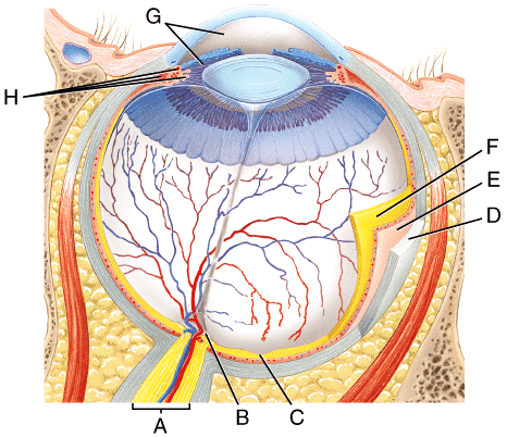
What is the choroid?
The choroid is the middle layer of tissue in the wall of the eye. It’s found between the sclera (the whites of the eyes) and the retina (the light-sensitive tissue in the back of the eye).
How thick is the choroid?
The thickness of the choroid varies depending on what part of the eye it’s lining. For example, it’s the thickest in the back of the eye (approximately 0.2 mm) and narrows to approximately 0.1mm as it gets to the peripheral part of the eyeball. There are four different layers of the choroid:
What is a benign brain tumor that develops in the choroid plexus?
Choroid plexus papilloma – Rare, benign brain tumor that develops in the choroid plexus (tissue that makes cerebrospinal fluid).
What is hemorrhagic choroidal detachment?
Hemorrhagic choroidal detachment – A “ hemorrhagic choroidal detachment ” occurs when blood fills the space between the sclera and choroid, such as when a blood vessel bursts. It is associated with high pressure in the eyes and can occur during surgery. It is usually more painful than a serous detachment.
What is a choroidal rupture?
Choroidal rupture – A tear in the choroid, Bruch’s membrane and the retinal pigment epithelium (RPE) that result from an eye injury. Choroid plexus papilloma – Rare, benign brain tumor that develops in the choroid plexus (tissue that makes cerebrospinal fluid).
What is the term for the separation of the choroid and sclera?
Choroidal detachment and hemorrhage – Separation of the choroid from the sclera; this may happen as a result of low eye pressure (serous choroidopathy, which is fluid-filled) or high eye pressure (hemorrhagic choroidopathy, which is blood-filled).
What is the term for inflammation of the choroid?
Chorioretinitis – Inflammation of the choroid caused by infection or an autoimmune disease. Choroideremia – A hereditary, progressive deterioration of the choroid; this condition primarily affects men.
What is the choroid plexus?
Tests. The choroid plexus is a thin structure that lines most of the the ventricles of the brain. It is a protective barrier that produces cerebrospinal fluid (CSF), a fluid that provides nourishment and cushioning for the brain and spinal cord. 1 . Cysts or tumors can form in the choroid plexus, and the cysts are not usually as dangerous as ...
Which part of the brain is directly adherent to the choroid plexus?
The pia mater and the choroid plexus are directly adherent to the brain tissue, while there is a small space between the brain and the other layers of the meninges (dura mater and arachnoid mater). The pia mater covers the whole CNS, but the choroid plexus is only present in some of the regions of the pia mater.
What are the structural issues that arise from the choroid plexus?
5 And a number of neurological conditions affect and are impacted by the choroid plexus and/or CSF flow. 1
What is the blood CSF barrier?
Blood-CSF barrier: The blood-CSF barrier, which is created by the choroid plexus and the meninges, helps protect the brain from infectious organisms and helps maintain control of the nourishment and waste in and out of the brain. 2 The permeability of this structure affects the ability of medications, drugs, and other substances to enter the brain.
What are the anatomical variations of the choroid plexus?
Anatomical Variations. Variations in the function or structure of the choroid plexus can be associated with cysts and other congenital (from birth) malformations. 3 If they block CSF flow, choroid plexus cysts can lead to hydrocephalus and other brain malformations.
Where does the choroid plexus flow?
The choroid plexus-produced CSF flows around the surface of the whole CNS.
Can choroid plexus cysts be detected?
There may be an increased incidence of choroid plexus cysts among newborns who have other birth defects. The cysts can often be detected before birth with a fetal ultrasound. 4
What is the choroid?
Choroid forms a vital structure of the eye which could be included in several pathologies. It is of immense significance provided its functions such as thermoregulation, vascularization and even in the production of the growth factors. Choroid, histologically, exhibits 5 layers – outer pigment layer, suprachoroid; two Vascular layers, Haller (external) and Sattler (internal), choriocapillaris layer and the Bruch’s membrane.
How many faces does the choroid have?
The choroid structure exhibits two faces – the internal is concave and accommodates the retina with no adherence while the external is convex, solidarized with sclera through the ciliary nerves, vessels and the lax connective tissue. The choroid comprises 2 openings – as a demarcation, an anterior one with ora serrata and the other as a posterior one which passes through the optic nerve.
What is the inflammation of the choroid that is a result of an autoimmune disease or an infection?
Chorioretinitis – it is the inflammation of the choroid which is as a result of an autoimmune disease or an infection
Why is choroidal circulation important?
The choroidal circulation is said to be responsible for about 85% of the blood flow in the eye, thus making it an important structure to the function of eyes. Some other functions are –
Which membrane forms the innermost layer of the choroid?
Bruch’s membrane – it forms the choroid’s innermost layer. This transparent layer imparts a homogeneous appearance indicated by an endothelial basement membrane from the choriocapillaris layer of the capillaries
What is the term for a tear in the choroid?
Choroidal rupture: the retinal pigment epithelium and Bruch’s membrane which lead to an eye injury, tear in the choroid
Where is the choroid plexus located?
The cerebral aqueduct is void of choroid plexus. The choroid plexus is located in the posterior medullary velum which partially forms the roof of the fourth ventricle.
When does the choroid plexus develop?
Signs of choroid plexus development of the fourth ventricle are evident around the 6th or 7th week of gestation , with the choroid plexus of the lateral ventricles developing at the same time, or shortly after in week 7. The choroid plexus of the third ventricle generally begins to develop a bit later in week 8.
What is the most common treatment for choroid plexus papilloma?
A solid mass, with some calcifications, is generally evident on imaging. The most common treatment for choroid plexus papilloma is complete surgical excision.
What type of cell is the choroid plexus?
a fourth ventricle. These ventricles are lined by a specialized type of glial cell called ependymal cells, or the ependyma. The choroid plexus is formed by these vascularized invaginations, bordered by the ependyma.
Where is choroid plexus papilloma most common?
This condition is most commonly seen in the lateral ventricle of children, with over 85% of cases occurring in children under the age of 5. Tumors can also develop in adulthood, however, they are most likely to develop in the fourth ventricle at this point.
Which artery supplies the choroid plexus?
The choroid plexus of the lateral ventricles are supplied by the anterior choroidal arteries (branch of internal carotid artery) and the lateral posterior choroidal arteries (branch of the posterior cerebral artery ). Anterior choroidal artery (caudal view)
Which ganglion controls blood flow to the choroid plexus?
The choroid plexus receives sympathetic and parasympathetic innervation. Sympathetic fibers from the superior cervical ganglion control blood flow to the choroid plexus, while parasympathetic fibers reduce CSF production.

Popular Posts:
- 1. which event shows how eckels character deloved over the course of the story
- 2. what does one learn in a marketing consumer behavior course
- 3. what is faster long course
- 4. ucsb how to update course units
- 5. why is my hair so course and braking?
- 6. how long does a paralegal course take
- 7. what does the course business principles look like
- 8. how to remove a course from your udemy list
- 9. how far is okaloosa island from timber creek golf course
- 10. how to contact city to design disc gold course